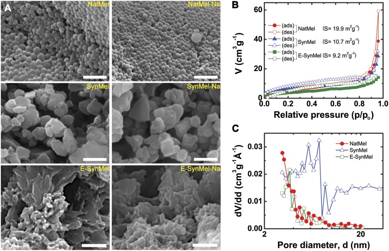
Fig. 1.
Structural characterization of eumelanins. (A) SEM images of pristine natural (NatMel), Na+-loaded natural (NatMel-Na), synthetic (SynMel), Na+-loaded synthetic (SynMel-Na), electro-deposited (E-SynMel), and Na+-loaded electrodeposited (E-SynMel-Na) melanins. Scale bar, 500 nm. (B) Nitrogen adsorption–desorption isotherms and (C) pore-size distributions as determined by using the Barrett–Joyner–Halenda method.

