Fig. 3.
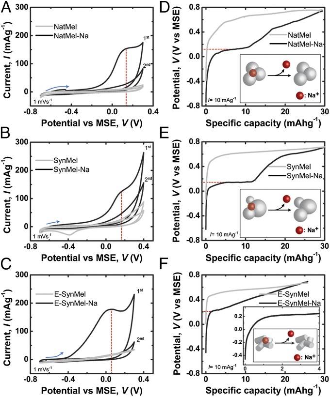
Electrochemical characterization of eumelanins. (A–C) CV of melanins in 1 M Na2SO4 electrolyte indicate redox peaks between 0 and ∼0.2 V (vs. MSE). Galvanostatic half-cell discharge profiles of melanins at 10 mAg−1 measured in 1 M Na2SO4 with Pt counter and MSE reference electrodes are shown in D–F. Plateaus at potentials of ∼0.2 V indicate the release of sodium ions from Na+-loaded melanin electrodes.

