Fig. 3.
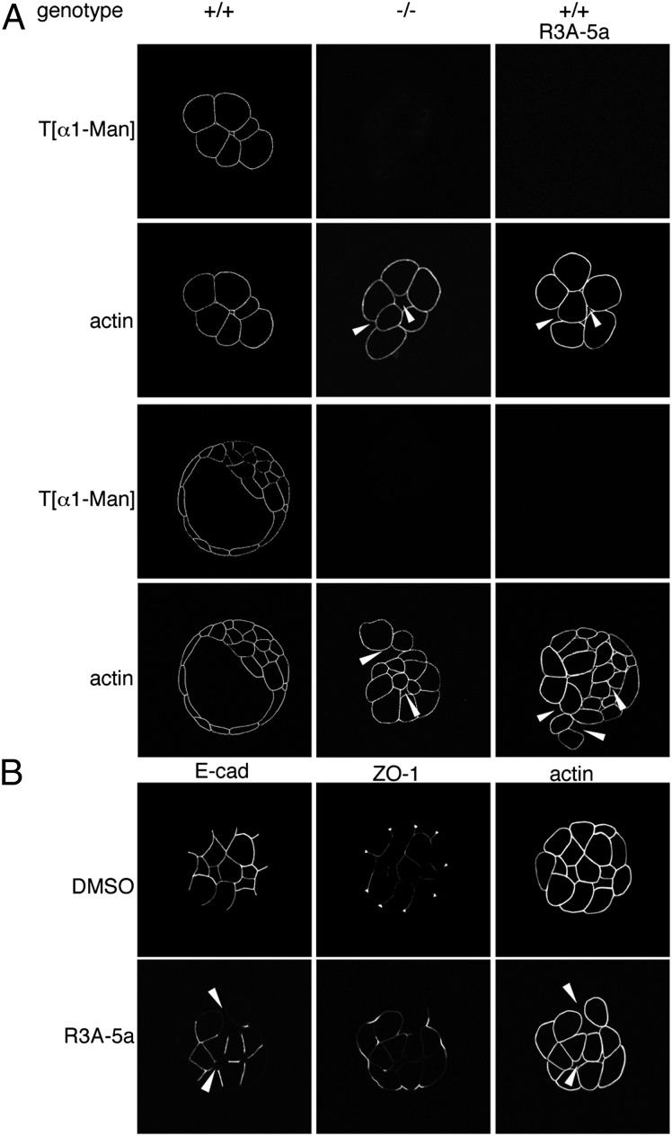
Characterization of O-mannosylation impaired embryos. (A) Whole-mount immunofluorescence analyses of embryos from intercrosses of Pomt2+/− mice and of WT embryos treated with R3A-5a. Morula- and blastocyst-stage embryos were analyzed with the T[α1-Man]–specific antibody and phalloidin. O-mannosyl glycans were not detectable in Pomt2−/− or inhibitor-treated embryos. Arrowheads indicate sites of impaired blastomere adhesion. Genotypes of the individual embryos shown were determined by PCR analysis as in Fig. 1, following microscopy. (B) Whole-mount immunofluorescent analysis of adherens and tight junctions in WT embryos after R3A-5a or mock (DMSO) treatment. Morula-stage embryos stained with E-cad– or ZO-1–directed antibodies or with phalloidin are shown. Arrowheads indicate diminished E-cad staining at sites of reduced blastomere attachment.

