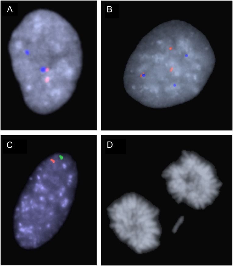
Fig. 2.
Examples of aneusomy and chromosomal mis-segregation. (A) Nucleus from case W1 with two signals from both chromosome 17 probes, corresponding to disomy. (B) Trisomy 17 in a cell from case D3, using the same probe combination as in A. (C) Monosomy 2 in a cell from case P1, using the chromosome 2 probe pair. (D) Lagging chromosome at telophase in triploidy case T1.

