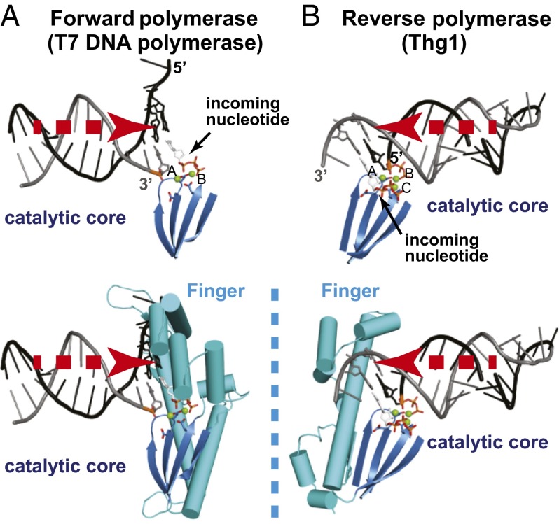
Fig. 1.
Reverse polymerization is a mirror image of forward polymerization. In forward and reverse polymerases, the approach of nucleic substrate binding is concurrent with the direction of polymerization. The catalytic core and the finger domain of the polymerases are shown as blue and cyan cartoon models, respectively. Stick models indicate specific base pairing and incoming nucleotides. Magnesium ions are shown as green spheres. (A) Direction of substrate binding and domain organization of T7 DNA polymerase as a forward polymerase. (B) The direction of substrate binding and domain organization of Thg1 as a reverse polymerase. The triphosphate moiety of the ppp-tRNA model was generated based on that of ATP in the CaThg1-ATP structure.

