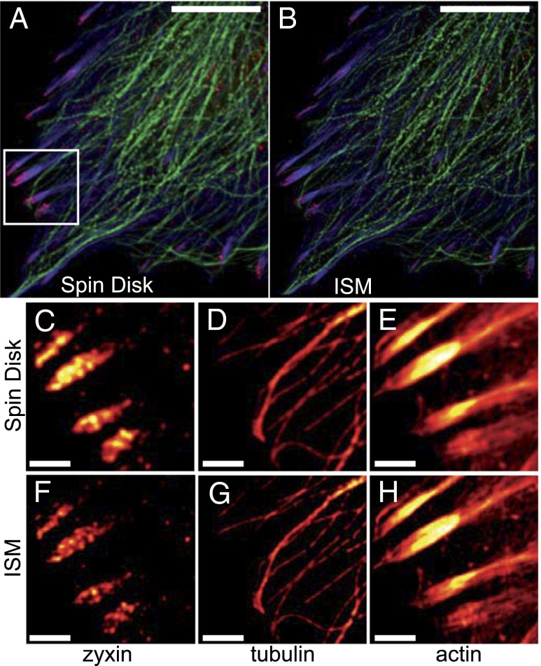
Fig. 5.
ISM allows higher resolution in multicolor imaging. Multicolor ISM of fixed REF52 cells showing Alexa 488-labeled actin (blue) and TRITC-labeled tubulin (green) cytoskeletal networks together with the Alexa 647-labeled focal adhesion protein zyxin (red). A confocal image is shown in A. The corresponding CSD-ISM image from 378 single images is shown in B. For CSD-ISM images of Alexa 488 and TRITC, each single image was exposed for 66 µs (∼15 s acquisition time), and for Alexa 647 an exposure of 210 µs (∼45 s acquisition time) was used. Magnifications of the indicated regions of interest are shown in C–H. The scale bars are 10 μm for the overview and 2 μm for the regions of interest.

