Fig. 6.
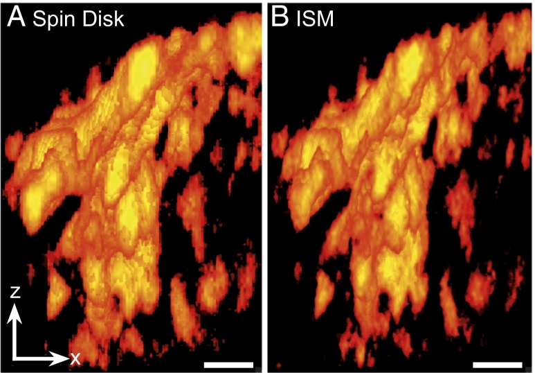
ISM gives enhanced resolution in three dimensions. Three-dimensional representation of confocal (A) and CSD-ISM (B) z-stack data of GFP-fused 3PO-Tau aggregates in an N1E115 neuroblastoma cell. The increased level of detail in the ISM data allows for better characterization of different types and progression of aggregates. The data were acquired using the 16-pulse laser sequence (Synchronization and Data Acquisition). The images were calculated from 125 single images with an exposure time of 6 µs per image (∼0.8 s acquisition time). The scale bar (1 μm) is the same for x- and z-direction. A movie of these data may be found as Movie S1.

