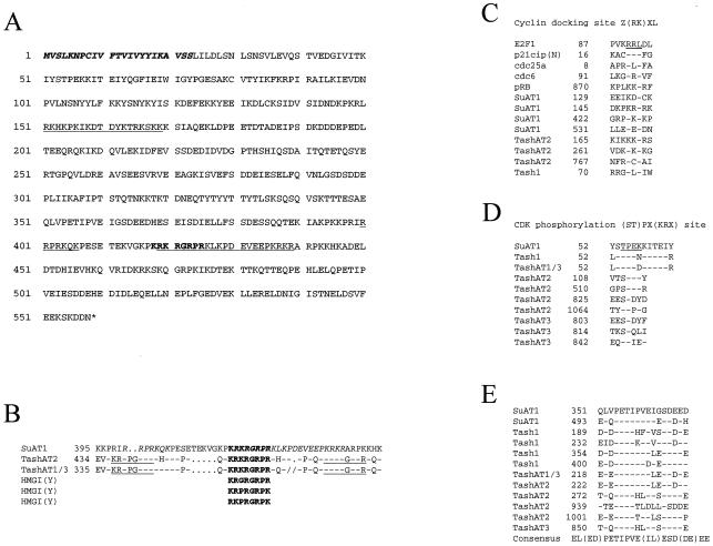
FIG. 2.
Identification of SuAT1 peptide motifs. (A) Predicted amino acid sequence of SuAT1. The putative signal sequence is in boldface italics, the conserved AT hook DNA binding domain is in boldface, and the three potential nuclear localization signals are underlined. (B) Comparison of the AT hook region of SuAT1 with those of TashAT2, TashAT1 and TashAT3 (TashAT1/3; regions 1 to 3), and the HMGI(Y) protein. The conserved AT hook motif is shown in boldface, and nuclear localization signals are in italics. (C) Comparison of potential SuAT1 cyclin A docking sites with related motifs of Tash1 and TashAT2 and the cell cycle polypeptides E2F1, p21cip, cdc25a, cdc6, and pRB. (D and E) Comparisons of CDK phosphorylation sites (D) and a repeated region of unknown function (E) with related motifs of Tash1, TashAT1, TashAT2, TashAT3, and TashAT1-TashAT3 (TashAT1/3).

