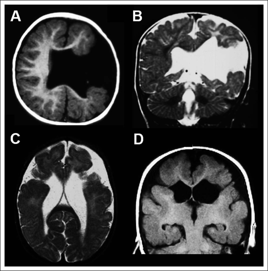
Figure 1.
Magnetic resonance imaging (MRI) appearance of schizencephaly. T1-weighted axial (A) and T2-weighted coronal (B) images from an 11-month-old boy with unilateral left hemisphere open-lip schizencephaly demonstrate a wide cleft lined by gray matter extending from the lateral ventricle to the pial surface. T2-weighted axial (C) and T1-weighted coronal (D) images from a 6-year-old girl with bilateral closed-lip schizencephaly demonstrate clefts extending from the lateral ventricles to the pial surface in both hemispheres; in this case, the regions of gray matter on either side of the clefts are closely apposed.

