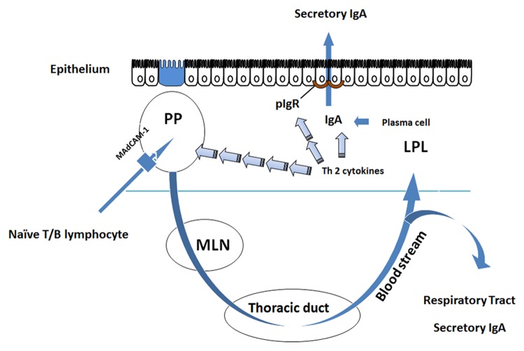
Figure 1. Adaptive mucosal immune function in the small intestine. Naïve T and B lymphocytes enter the Peyer patches (PP) via interaction with their integrins L-selectin or α4β7 and MAdCAM-1 which is present on the high endothelial venules (HEV) of the PP. The cells are sampled and sensitized to antigen in Microfold (M) cells inside the PP. Sensitized cells return to the peripheral circulation via the thoracic duct and are distributed to other mucosal effector sites. Within these effector sites, Th2 cytokines (IL-4, IL-5, IL-6, and IL-10) produced by T lymphocytes stimulate maturation of B cells into competent IgA producing cells, i.e., plasma cells. These cells produce the principal molecule of adaptive immunity, dimeric IgA, in the lamina propria where it undergoes transepithelial transport via polymeric immunoglobulin receptor (pIgR) which is expressed on the basal surface of mucosal enterocytes.

An official website of the United States government
Here's how you know
Official websites use .gov
A
.gov website belongs to an official
government organization in the United States.
Secure .gov websites use HTTPS
A lock (
) or https:// means you've safely
connected to the .gov website. Share sensitive
information only on official, secure websites.
