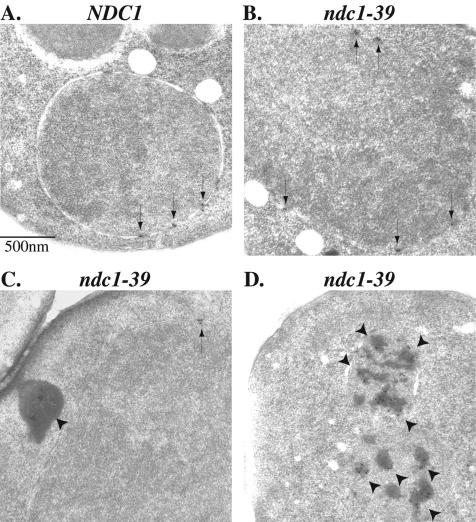
FIG. 8.
IEM of Nup49p-GFP assembly in NDC1 and ndc1-39 cells at the restrictive temperature. Experiments were performed as described for Fig. 7. Nup49p-GFP was immunolabeled with an antibody to GFP and visualized with a colloidal gold-conjugated secondary antibody in NDC1 (A) and ndc1-39 (B to D) cells after the repression of Nup49p-GFP expression in YPD medium. At 35°C, Nup49p-GFP was localized correctly to the nuclear pores (arrows) in NDC1 cells (A) and in some of the ndc1-39 cells (B and C). Nup49p-GFP labeling was also observed seven times as aggregates in the cytoplasm (arrowheads in panel C) and four times as membrane-bound aggregates (arrowheads in panel D) in the 15 ndc1-39 cells examined. Bar, 0.5 μm.

