
Figure 1.
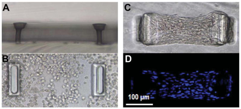
A) micro-tissue gauge (μTUG) well shown A) from the side without cells and gel, B) from the top filled with a cell-populated gel at t=1 hours and C & D) at t=72 hours. In (D) the nuclei are stained by Hoechst to facilitate cell counting utilizing fluorescent microscopy. All panels imaged at 20X.