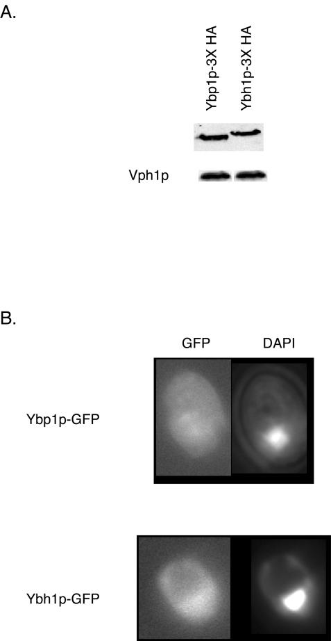
FIG. 4.
Expression and localization of Ybp1p and Ybh1p. (A) Whole-cell protein extracts were prepared from strains expressing 3× HA-tagged forms of Ybp1p or Ybh1p. Equal amounts of protein were electrophoresed by SDS-PAGE and subjected to Western blot analysis with anti-HA antibody to detect the epitope-tagged proteins. The blot was also probed with antibody directed against the vacuolar ATPase subunit Vph1p to ensure equal protein loading. (B) Strains containing integrated Ybp1p- or Ybh1p-GFP fusion genes were grown to mid-log phase, stained with DAPI, and visualized by fluorescence microscopy.

