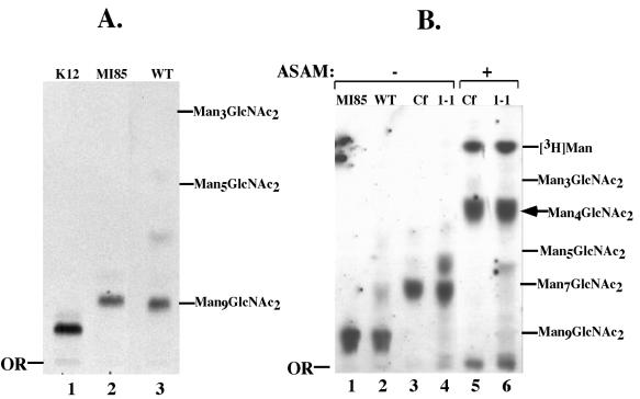
FIG. 5.
Structural characterization of OSL oligosaccharides. [3H]Man-labeled glycans were analyzed by TLC and autoradiography. (A) Lane 1, 3H-labeled Glc3Man9GlcNAc2 from WT CHO cells (K12); lane 2, 3H-labeled Man9GlcNAc2 from MI8-5 mutant CHO cells; lane 3, 3H-labeled OSL glycan from WT T. brucei. (B) 3H-labeled OSL glycans from MI8-5 CHO cells (lane 1), WT T. brucei (lane 2), C. fasciculata (lanes 3 and 5), and ConA 1-1 (lanes 4 and 6). Glycans in lanes 5 and 6 were treated with A. saitoi α-mannosidase (ASAM), and those in lanes 3 and 4 were mock treated. Positions of glycan standards (2 μg; Sigma) visualized by orcinol-H2SO4 staining are indicated on the right. Cf, C. fasciculata glycan.

