
Figure 7.
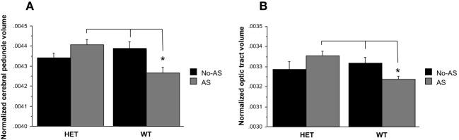
Analysis of the normalized volumes of 62 structures showed that a significant effect of AS in two white matter regions: the cerebral peduncle (10% FDR) and the optic tract (5% FDR). WT-AS mice showed a smaller cerebral peduncle (A) and optic tract (B) volumes relative to WT-No-AS mice and to HET-AS mice (*p < 0.02). Results are reported as mean ± s.e.m.