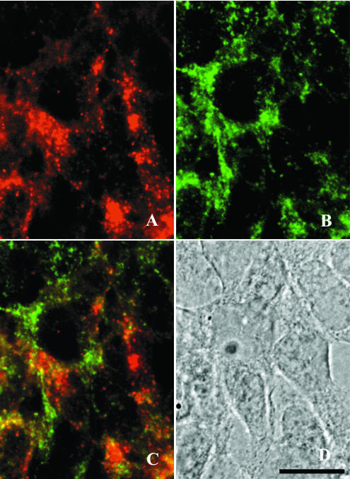
FIG. 5.
Double immunolabeling of (A) PrP and (B) LAMP-1 in ScGT1-1 cells treated with E-64d. Cells were exposed to guanidine thiocyanate prior to immunolabeling. A merged image of the red and green channels shows partial colocalization of PrP and LAMP-1 (C). A bright-field micrograph of the cells is also shown (D). Bar, 20 μm.

