
Figure 1.
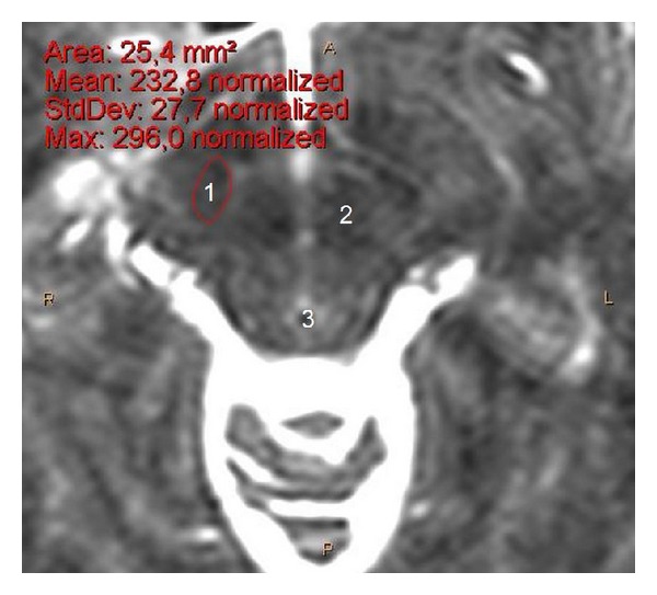
Transverse cerebral MRI section (T2-weighted, 1.5 Tesla) at level Z = −4 (zoom on mesencephalon), from a 37-year-old male, showing the STN location and surface area. 1: STN, 2: red nucleus, 3: aqueduct (of Sylvius), A: anterior, P: posterior and, R: right, L left.