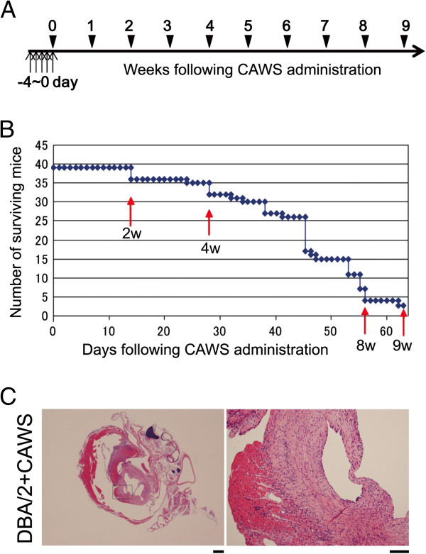
Figure 1.
Survival curve and histopathological analysis. (A) Schedule of CAWS administration. Vertical arrows indicate that CAWS was administered i.p. (0 or 1 mg/mouse) for 5 consecutive days to each B6 or DBA/2 mouse. (B) The surviving number of DBA/2 mice following CAWS administration is shown, and confirms that CAWS administration is as effective (toxic) as in our previous report [12]. Red vertical arrows indicate the time points (2 w, 4 w, 8 w and 9 w) at which three B6 mice and three DBA/2 mice were sacrificed for mRNA purification. (C) Histopathological analysis was performed on aortic roots and coronary arteries collected from DBA/2 mice at 6 w following CAWS administration stained with H&E. Enlarged views of the region indicated by rectangles in the left panels are shown in the right panels. Bar = 500 μm for left panels and 100 μm for right panels.

