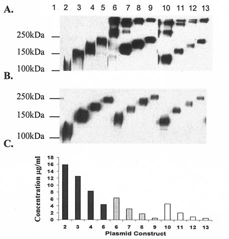
FIG. 2.
Expression of vaccine plasmids was visualized by Western blot analysis. Nonreducing (A) and reducing (B) SDS-PAGE analysis demonstrated that all constructs are efficiently expressed in vitro and FT-stabilized versions of sgp140YU-2(−) form trimers even after boiling and under denaturing conditions with and without the addition of one, two, or three copies of mC3d. Human embryonic kidney cells were transiently transfected with 2 μg of plasmid DNA from each vaccine plasmid. Supernatants were collected, and 1.5% of the total volume was subjected to electrophoresis. Each blot was probed with HIV-Ig followed by anti-human IgG conjugated to horseradish peroxidase and detected with enhanced chemiluminescence. (C) Expression ELISA data to determine the relative level of expression of codon-optimized DNA vaccine plasmids were quantified using 1.5% of the total supernatant from transiently transfected 293T cells and comparing relative amounts to recombinant gp120YU-2 (ImmunoDiagnostics) as a standard and are expressed as micrograms per milliliter of Env secreted into the supernatant. Lane 1, molecular mass marker; lane 2, sgp120YU-2; lane 3, sgp120YU-2 mC3d1; lane 4, sgp120YU-2-mC3d2; lane 5, sgp120YU-2-mC3d3; lane 6, sgp140YU-2(−); lane 7, sgp140YU-2(−)-mC3d1; lane 8, sgp140YU-2(−)-mC3d2; lane 9, sgp140YU-2(−)-mC3d3; lane 10, sgp140YU-2(−/FT); lane 11, sgp140YU-2(−/FT)-mC3d1; lane 12, sgp140YU-2(−/FT)-mC3d2; lane 13, sgp140YU-2(−/FT)-mC3d3.

