
Figure 6.
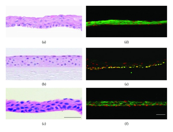
Histological and immunohistochemical analyses of cell sheets. HE staining was performed for an oral mucosal epithelial cell sheet (a), a normal cornea (b), and a corneal epithelial cell sheet (c). Human oral mucosal epithelial cell sheets were stained with antikeratin 3/76 (d), anti-p63 (e), and anti-ZO-1 (f) antibodies. Nuclei were costained with Hoechst 33342. Scale bars: 50 μm.