
Figure 9.
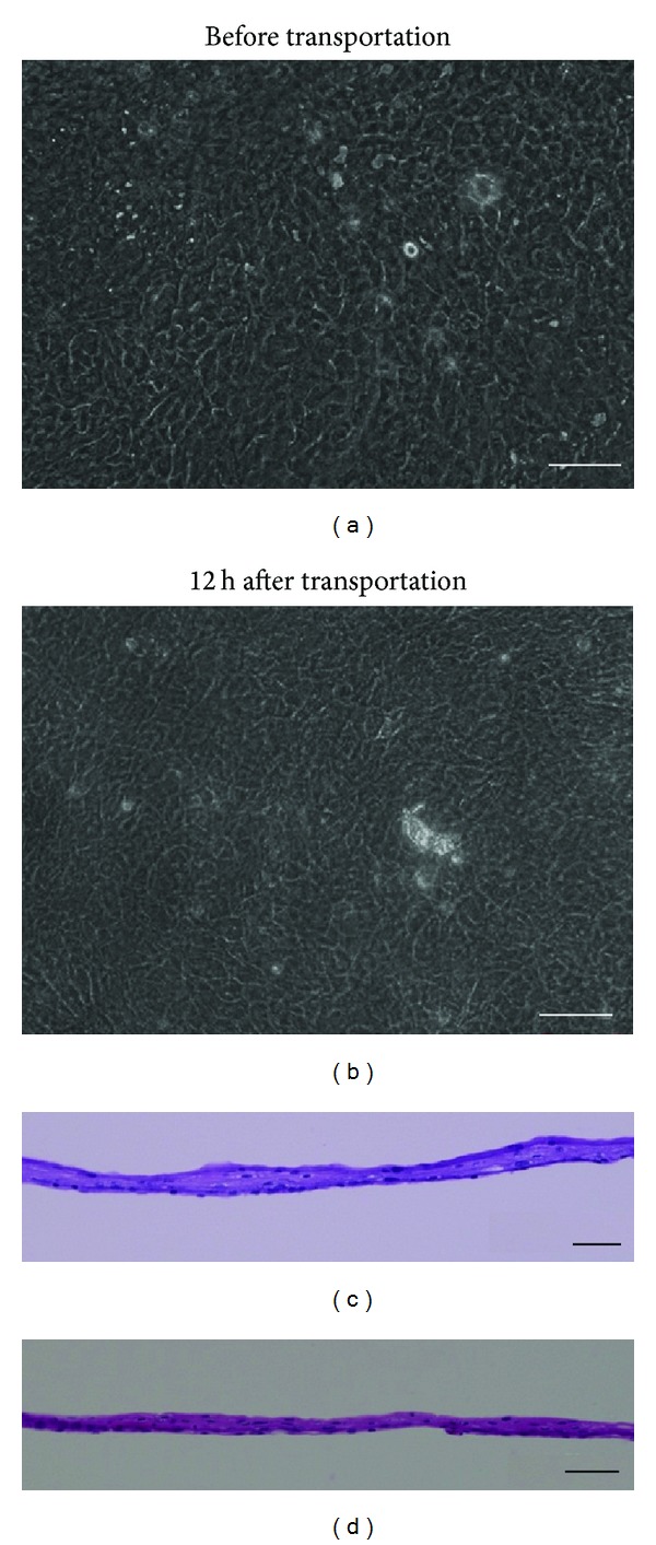
Human oral mucosal epithelial cell sheets before and 12 h after transportation. The cell morphology was examined using phase-contrast microscopy (a, b) and HE staining (c, d). Scale bars: 100 μm (a, b), 50 μm (c, d).
Official websites use .gov
A
.gov website belongs to an official
government organization in the United States.
Secure .gov websites use HTTPS
A lock (
) or https:// means you've safely
connected to the .gov website. Share sensitive
information only on official, secure websites.
Human oral mucosal epithelial cell sheets before and 12 h after transportation. The cell morphology was examined using phase-contrast microscopy (a, b) and HE staining (c, d). Scale bars: 100 μm (a, b), 50 μm (c, d).