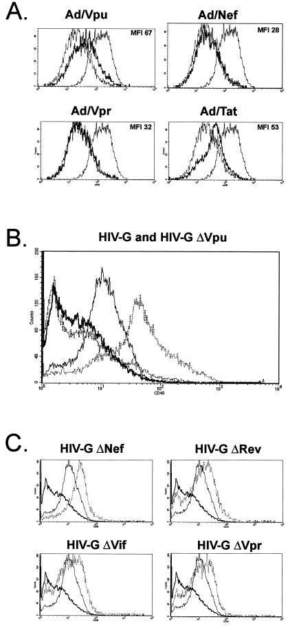
FIG. 2.
HIV-1 Vpu upregulates surface CD40 on endothelial cells. (A) HUVEC were infected with recombinant adenoviruses for 48 h, and quantitative CD40 expression was determined by flow cytometry. HUVEC infected with Ad/trans alone or stimulated with TNF-α (10 ng/ml) served as negative or positive controls for CD40 expression, respectively. For each panel, the profile obtained with the respective recombinant adenovirus is shown as a bold solid line. Superimposed are the baseline expression levels obtained with the Ad/trans control (dashed line) and the elevated expression levels induced by TNF-α-treatment (thin solid line). The MFI value for each profile is given in parentheses. An increase in CD40 expression was detected on cells infected with Ad/Vpu (MFI 67) relative to cells infected with Ad/trans alone (MFI 37). No change was seen in cells infected with Ad/Nef (MFI 28), Ad/Vpr (MFI 32), Ad/Rev (not shown), or Ad/Vif (not shown). A moderate increase in CD40 was detected in cells infected with Ad/Tat (MFI 53). CD40 expression was efficiently induced by TNF-α (MFI 88). (B) HUVEC were infected with an HIV-1 pseudotyped virus (HIV-G) (thin solid line), as well as with an HIV-Gmutant lacking Vpu (ΔVpu) (bold solid line), and the expression of surface CD40 was evaluated by flow cytometry. Mock-infected (bold hatched line) and TNF-α-stimulated (thin hatched line) HUVEC served as negative and positive controls for CD40 expression, respectively. Constitutive CD40 levels on mock-infected EC were low (MFI 2.7) but were significantly increased by infection with HIV-G (MFI 10.4). Infection of EC with the ΔVpu mutant did not lead to CD40 induction (MFI 2.8). CD40 expression was efficiently induced by TNF-α (MFI 32.5). (C) Expression of CD40 on HUVEC infected with HIV-G mutants (hatched lines) lacking HIV-1 Nef (ΔNef), Rev (ΔRev), Vif (ΔVif), or Vpr (ΔVpr) was similar to that of the wild-type HIV-G (thin solid line). The decrease in CD40 seen with ΔVpu HIV-G (bold line) is superimposed on each image for reference.

