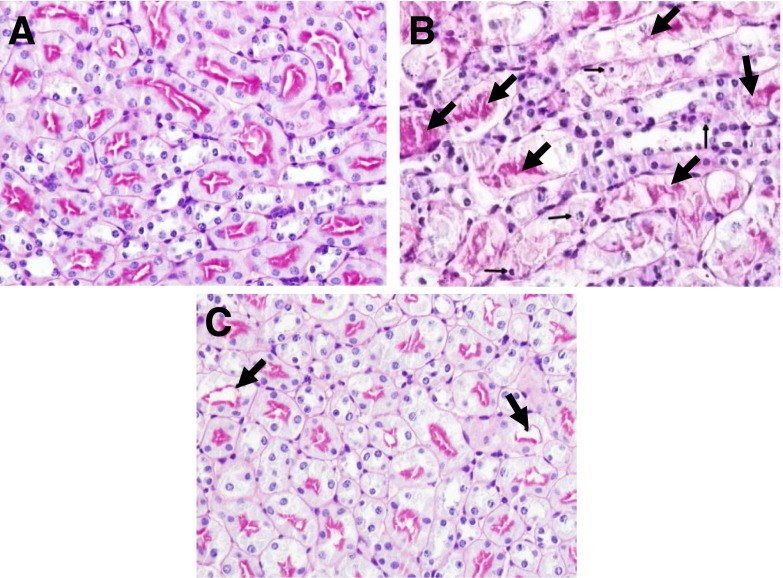
Fig. 6.
Representative pictures of kidney histology. PAS–stained section of (A) wild-type sham-operated, (B) wild-type ischemic AKI, and (C) NLRP3−/− mouse ischemic AKI are shown. In wild-type sham-operated (A) kidneys, the tubules have well-defined brush borders without necrosis or apoptosis. In wild-type ischemic AKI (B) there is acute tubular necrosis (large arrows) and tubular cell apoptosis (small arrows). In NLRP3−/− mouse ischemic AKI (C), acute tubular necrosis and tubular cell apoptosis is statistically significantly reduced.

