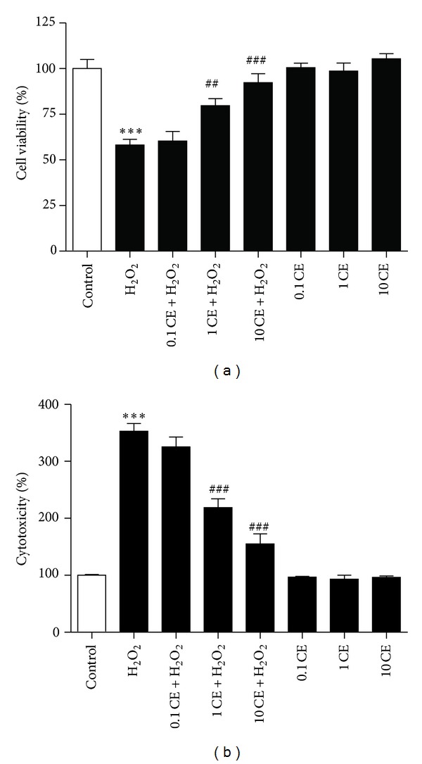
Figure 1.

Protective effects of CE against oxidative stress in primary cultured cortical neurons. (a) Bar graphs showing the reduced cell viability after H2O2 treatment that was significantly attenuated by the treatment of the neurons with 1 mg/mL and 10 mg/mL CE as tested by MTT assay. (b) Bar graphs showing the H2O2 induced cytotoxicity that was significantly reduced by the treatment of the neurons with 1 mg/mL and 10 mg/mL CE as tested by LDH release assay. ∗ refers to a comparison of the control group and # refers to a comparison of the H2O2 group.
