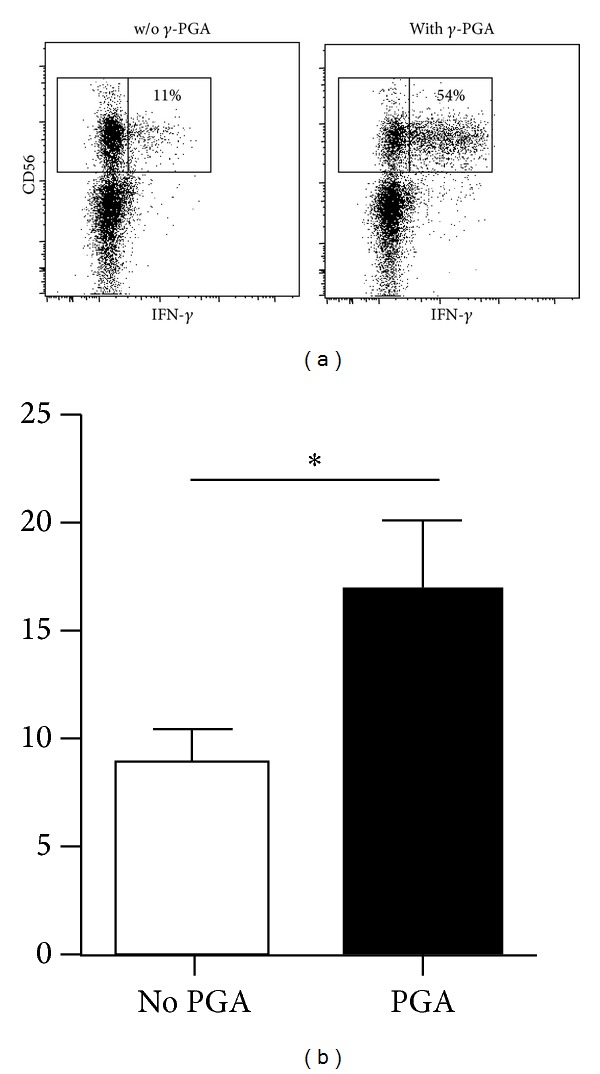
Figure 5.

PBMCs from healthy donors were incubated with or without γ-PGA for 8 hours and then stimulated with K562 cells for an additional 7 hours. (a) Dot plots show CD56+CD3− NK cells stained for intracellular IFN-γ from a representative donor. Inset numbers indicate the percentage of NK cells that produced IFN-γ. (b) Bar graph shows the average percentage of IFN-γ + NK cells from 16 donors; *P < 0.05.
