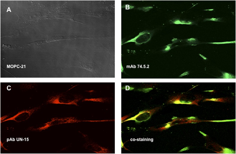
FIGURE 1.
Colocalization of the mature and full-length gC1qR on the cell surface of ECs. Human brain microvascular cells were grown on glass cover slips, and the attached monolayer of cells were incubated with PBS containing 0.1% BSA and 1 mg/ml Fc fragments to block FcRs, followed by incubation with MOPC-21 (A), mAb 74.5.2, or pAb UN15. The bound Abs were probed with either Alexa Fluor 488–F(ab′)2 anti-mouse Abs (B) or Alexa Fluor 594–F(ab′)2 anti-rabbit Abs (C). (D) The merged picture shows staining with mAb 74.5.2 and pAb UN15. Original magnification ×68. The experiment is a representative of three such experiments. The control staining with rabbit nonimmune IgG, was similar to that in (A) and is not included.

