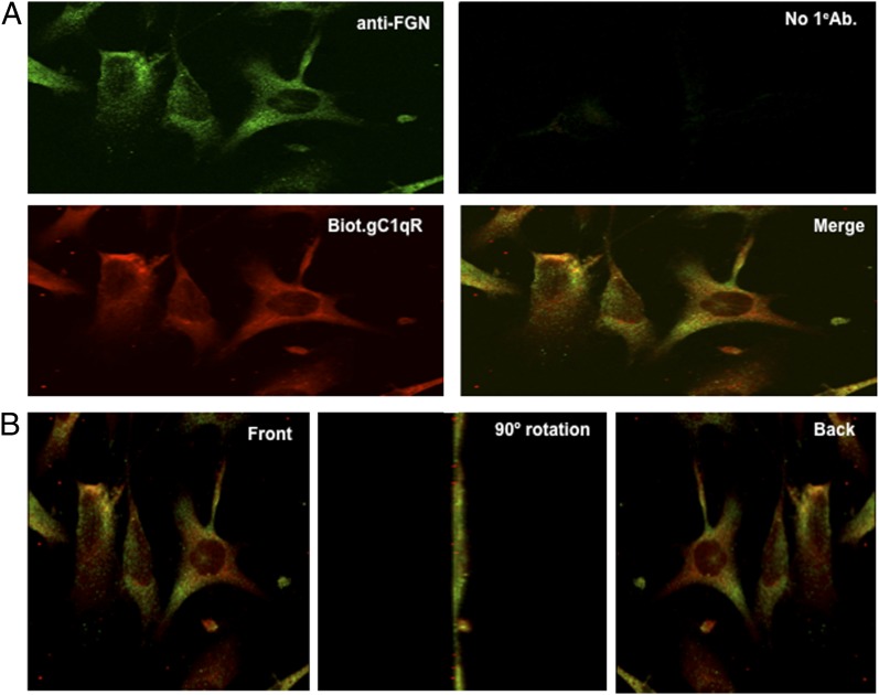
FIGURE 8.
Colocalization of gC1qR and FGN on the EC surface. (A) ECs were grown on cover slips and incubated with anti-FGN, biotinylated-gC1qR (Biot.gC1qR), or both (merge). Cells incubated without primary Ab (No 1eAb) were used as negative control. (B) Cells showing colocalization of FGN and gC1qR (merge) were subjected to three-dimensional rotation to show staining of cells as seen from the front (or top), the side (90° rotation), or back (360° rotation). Original magnification ×68.

