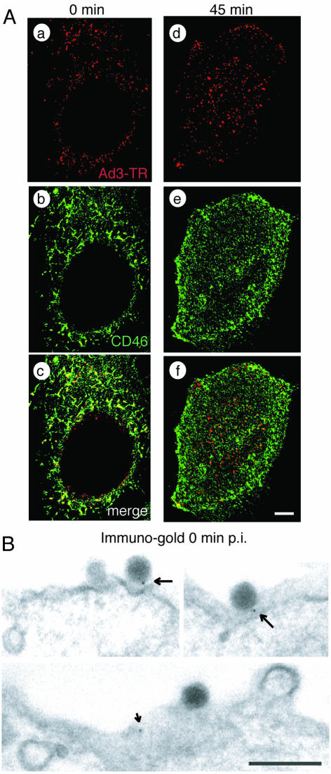
FIG. 4.
Ad3 colocalizes with cell surface CD46 of HeLa cells. (A) CLSM analyses of TR-labeled Ad3 (red) and CD46 stained with non-function-blocking antibody MCI20.6 (green). Single sections are shown at 0 min (a to c) and at 45 min (d to f) p.i. Scale bar, 5 μm. (B) Transmission EM showing immunogold staining of CD46 at 0 min p.i. Arrows indicate Ad3 associated with protein A-gold directed to anti-CD46; the small arrow indicates a gold particle not associated with Ad3. Scale bar, 200 nm.

