
Figure 7. MRI molecular imaging of inflammation in abdominal aortic aneurysm (AAA).
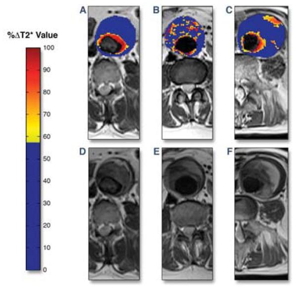
Representative color maps of T2-weighted MRI signal change (blue=low, red=high) induced by local accumulation of ultrasmall superparamagnetic particles of iron oxide (USPIO) in 3 patients with AAA. (A) A patient with enhanced periluminal T2 signal. (B) More diffuse, non-contiguous T2 changes present within the intraluminal thrombus, but not involving the aortic wall. (C) Periluminal T2 signal change with focal aortic T2 enhancement revealing USPIO accumulation and inflammation within the AAA wall. (D-E) T2-weighted MRI images without the superimposed color maps for comparison demonstrating focal T2 signal loss at USPIO-rich sites. Reproduced with permission from (32).