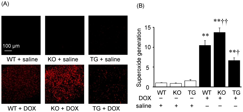
Figure 3. DOX-induced myocardial oxidative stress in WT, SMP30 KO and SMP30 TG mice.
A, Photomicrograph of DOX-induced superoxide formation from frozen heart sections using dihydroethidium (DHE). B, The mean of DHE fluorescence intensity of cardiomyocytes. Data were obtained from 8 mice in each group. **P<0.01 vs. same genotype mice given saline, †P<0.05 and ††P<0.01 vs. the doxorubicin-infused WT mice.

