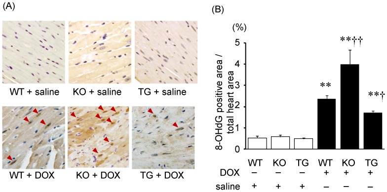
Figure 4. Immunohistochemical detection of oxidative DNA damage with 8-OHdG.
A, Photomicrographs of the 8-OHdG positive nuclei, which were stained with dark brown, prepared from heart specimens. B, 8-OHdG positive nuclei/total cells (%). Data were obtained from 7 mice in each group. **P<0.01 vs. same genotype mice given saline, †P<0.05 and ††P<0.01 vs. the doxorubicin-infused WT mice.

