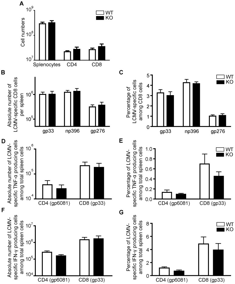
Figure 6. Normal in vivo anti-LCMV immune responses of STRA6 KO mice.
A. Spleen cell numbers on day 8 after LCMV infection. Means ± SD of absolute numbers of total splenocytes, CD4+ cells, and CD8+ cells in spleens of WT littermate control (n = 4) and KO (n = 4) mice on day 8 post-LCMV infection are presented. B and C. LCMV-specific CD8 cells on day 8 post-LCMV infection. On day 8 post-infection, the absolute numbers of gp33, np396 and gp276 tetramer-positive CD8 T cells per spleen (B) and the percentage of gp33, np396 and gp276 tetramer-positive cells among CD8 cells (C) were measured by flow cytometry. Means ± SD of data from 4 pairs of WT littermate control and STRA6 KO mice are presented. D and E. LCMV-specific TNF-α-producing CD4 and CD8 cells on day 8 post-LCMV infection. The absolute number of TNF-α-producing LCMV-specific CD4 cells (gp61-specific) and CD8 cells (gp33-specific) per spleen (D) and percentage (E) of these cells among total spleen cells of KO and WT mice on day 8 post-LCMV infection. Means ± SD of data from 4 pairs of STRA6 KO mice and WT littermate controls are shown. F and G. Virus-specific IFN-γ-producing CD4 and CD8 cells on day 8 post-LCMV infection. The absolute number of TNF-α-producing LCMV-specific CD4 cells (gp61-specific) and CD8 cells (gp33-specific) per spleen (D) and percentage (E) of these cells among total spleen cells of KO and WT mice on day 8 post-LCMV infection. Means ± SD of data from 4 pairs of STRA6 KO mice and WT littermate controls are shown. The results in this figure are analyzed by Student's t test. No significant difference was found between WT and groups.

