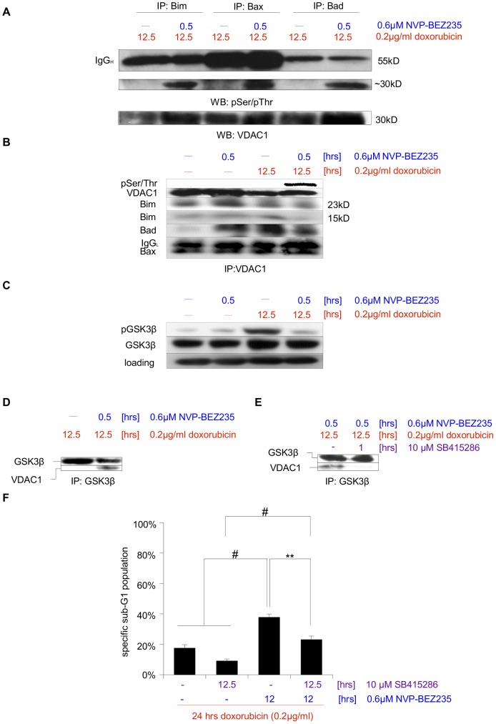
Figure 5. Sensitization for doxorubicin-induced apoptosis via posttreatment with NVP-BEZ235 is mediated via VDAC1.
A SHEP NB cells were treated for 12.5-BEZ235 for the last 0.5 hr. Either Bim, Bax or Bad was then immunoprecipitated and interaction partners that are phosphorylated on Serine or Threonine were visualized by Western blot analysis. A ∼30 kD protein, the presence of which appears to depend on NVP-BEZ235 addition, was identified as VDAC by VDAC1/Porin-specific antibody. IgGH – heavy chain. B Cells were left untreated, treated for 12.5 hrs with Doxorubicin, or after 12 hrs for 0.5 hr with NVP-BEZ235, or a combination of both (first 12 hrs with Doxorubicin alone, followed by the addition of NVP-BEZ235 for 0.5 hr). VDAC was immunoprecipitated and its phosphorylation status was probed. IgGL – light chain. C Cells were left untreated, treated for 12.5 hrs with doxorubicin, or after 12 hrs for 0.5 hr with NVP-BEZ235, or a combination of both (first 12 hrs with doxorubicin alone, followed by the addition of NVP-BEZ235 for 0.5 hr). Protein expression levels and phosphorylation status of GSK3β were analyzed by Western blotting, GAPDH served as loading control. D Cells were treated either for 12.5 hrs with doxorubicin, or a combination of doxorubicin and NVP-BEZ235, (first 12 hrs with doxorubicin alone, followed by the addition of NVP-BEZ235 for 0.5 hr). This was followed by immunoprecipitation of GSK3β and analysis of this protein's interaction with VDAC via immunoblotting. E Cells were again treated with a combination of doxorubicin and NVP-BEZ235, (first 12 hrs with doxorubicin alone, followed by the addition of NVP-BEZ235 for 0.5 hr), during the last hour in the absence or presence of the GSK3β-specific inhibitor SB415286. This was followed by immunoprecipitation of GSK3β and analysis of this protein's interaction with VDAC. F Apoptosis in cells treated for 24 hrs with doxorubicin, for 12.5 hrs with SB415286, for 12 hrs with NVP-BEZ235, or a combination of those substances was determined by FACS analysis of the DNA fragmentation of propidium iodide-stained nuclei, and percentage of specific DNA fragmentation is shown. Shown in A to E are representative blots of at least two independent experiments, in F the mean+s.e.m. of three independent experiments performed in triplicate is depicted. Statistical analysis was carried out by two-sided Student's t-test; * P-value <0.01; ** P-value <0.001; # P-value <0.0001.

