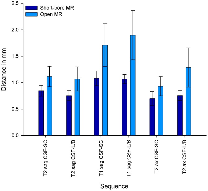
Figure 6. Contour Sharpness in MR images of the Cervicothoracic Spine.
Contour sharpness as distance in mm (±SD) that is neeeded for the signal to increase from 25% to 75% of the grayscale pixel value profile obtained with imageJ (see Figure S1). Contour sharpness was defined for the interface between corticospinal fluid (CSF) and spinal cord (SC), and between corticospinal fluid and vertebral body (B)/posterior longitudinal ligament (L) in sagittal T2-weighted, sagittal T1-weighted and axial T2-weighted sequences. P values were <0.0001 for all assessed contours.

