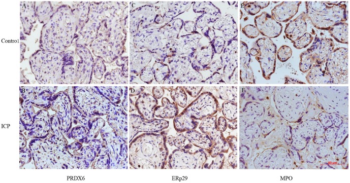
Figure 4. Immunohistochemical staining for PRDX6, ERp29 and MPO in the placental tissue from pregnant women with ICP and healthy pregnant women (×400).
Immunohistochemstry images demonstrated higher expression of PRDX6 (B) and ERp29 (D), and lower expression of MPO (F) in cytoplasm and/or nucleus of trophoblasts in the placenta from pregnant women with ICP than those in placenta from healthy pregnant women (A, C, E).

