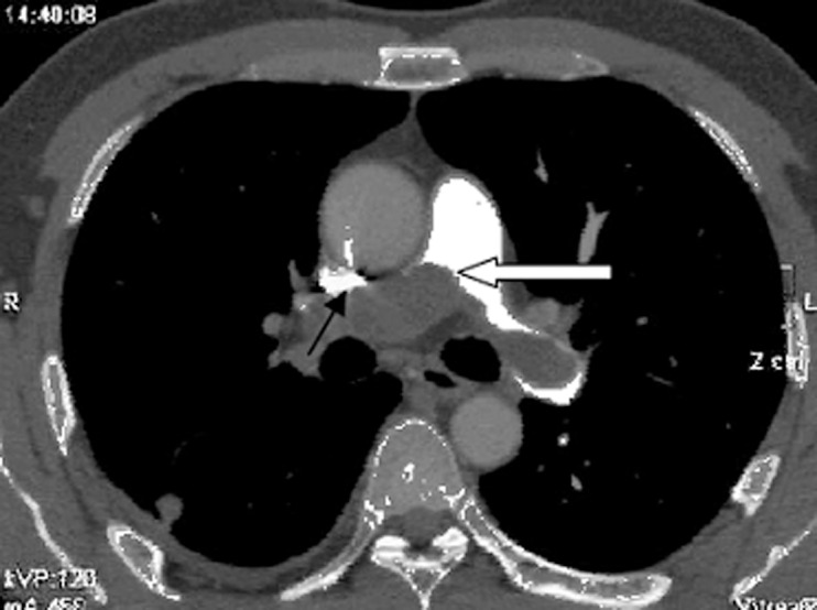
Figure 1. Pulmonary artery computed tomography angiography, axial view.

The lumen of the pulmonary trunk is obliterated by a low-density mass (white arrow) that extends into the left and right pulmonary arteries. Both walls of the left pulmonary artery are eclipsed by the lesion (black arrow). The pulmonary trunk, left pulmonary artery, and right pulmonary artery are fully or partially occupied by the lesion, and the proximal end of the lesion protrudes towards the right ventricular outflow tract (white arrow). We termed this appearance the wall eclipsing sign.
