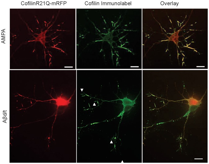
Figure 4. CofilinR21Q-mRFP incorporates into virtually all AMPA-induced rods, but only into half of rods in Aβd/t-treated neuronal cultures.
CofilinR21Q-mRFP fluorescence image, immunolabeled (Alexa 488) image, and overlay in neurons treated 30 min with 150 μM AMPA showing virtually all rods (immunolabel) have incorporated cofilinR21Q-mRFP. Similar results (not shown) were obtained for ATP-depleted neurons. CofilinR21Q-mRFP fluorescence image, immunolabeled image (Alexa 647 but colorized green), and overlay in neurons treated 24 h with Aβd/t. Immunolabeled rods that incorporated cofilinR21Q-mRFP were quantified from many different cultures and co-labeled rods accounted for 48±4% (std. deviation) of the total rods. Immunolabeled rods that do not contain mRFP are shown by arrowheads. Scale bars = 10 μm.

