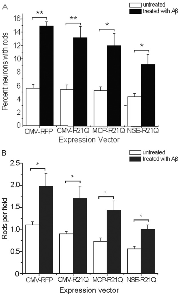
Figure 5. CofilinR21Q-mRFP reports on rod formation in hippocampal neurons in response to Aβd/t treatment.

(A) The fraction of cofilinR21Q-mRFP expressing neurons that formed rods after treatment with Aβd/t is 2 to 3 fold higher than for untreated neurons, regardless of which promoter drives expression, although more rods are detected when cofilin-R21Q-mRFP expression is greatest. (B) The number of rods per field in Aβd/t treated neurons expressing cofilinR21Q-mRFP is about 2 fold higher than the corresponding non-Aβd/t treated controls regardless of which promoter drives expression. (*Significant at p<0.05, **Significant at p<0.005, compared to their appropriate non-Aβd/t-treated control group).
