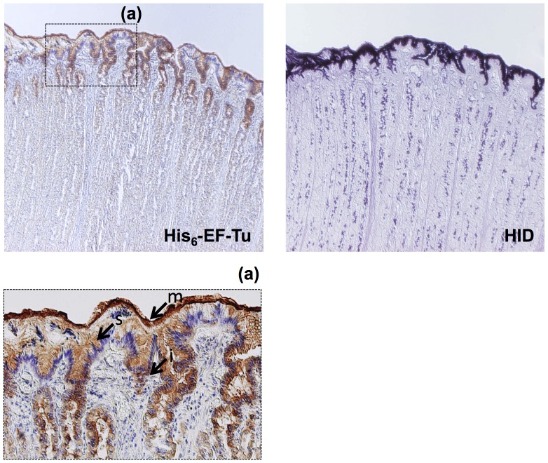
Figure 5. Histochemical staining of the porcine mucosal surface with His6-EF-Tu.
Binding of His6-EF-Tu was observed on the fixed (methanol-Carnoy) mucosal surface (right panels). These areas were coincident with areas positively stained for HID (left panel). Insets (a) are higher magnifications (200×) of the areas indicated by squares in the reference micrographs (40×magnification). Arrows indicate each region: mucous gel layer (m), surface mucous cells (s), and mucous cells around the isthmus region (i).

