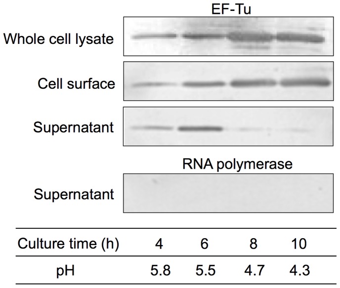
Figure 6. Localization of EF-Tu in L. reuteri JCM1081.
Supernatant, cell surface, and whole cell lysate fractions at different culture times were analyzed by western blotting. Outer surface proteins of L. reuteri JCM1081 were extracted with GHCl. Reactivity with anti-EF-Tu and anti-RNA polymerase antibodies is shown. An anti-RNA polymerase antibody was used to confirm whether cell lysis occurred. Culture time and pH are indicated in the table.

