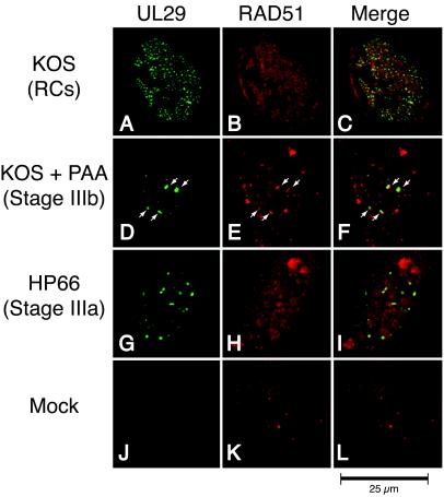
FIG. 4.
RAD51 colocalizes predominantly with UL29 in replication compartments (RCs) and stage IIIb foci but not stage IIIa foci. Vero cells were infected with KOS in either the absence (A to C) or presence (D to F) of PAA or infected with the polymerase null mutant virus HP66 (G to I). As described in Materials and Methods, cells were double labeled with mouse anti-UL29 (39S) and rabbit anti-RAD51 to detect the localization of the HSV-1 major DNA-binding protein UL29 (green) and cellular RAD51 (red). Viral structures examined included replication compartments, stage IIIb foci, and stage IIIa foci. Mock infection in the presence of PAA gave staining patterns similar to that of mock infection in the absence of PAA (J to L and results not shown).

