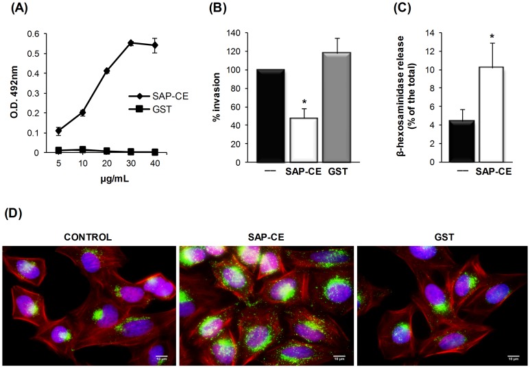
Figure 5. Cell adhesion and lysosome exocytosis-inducing properties of SAP-CE associated with T. cruzi metacyclic trypomastigote internalization.
(A) Increasing amounts of the purified recombinant protein SAP-CE or GST were added to 96-well plates covered with HeLa cells. After fixation and washes in PBS, the cells were incubated with MAb-SAP (diluted 1∶100) and with anti-mouse IgG peroxidase conjugate. The bound protein was revealed by o-phenylenediamine. Values are the means ± standard deviations of triplicates. (B) HeLa cells were incubated for 30 min with or without the recombinant protein SAP-CE or GST (40 μg/mL) and then incubated with metacyclic forms. After incubation for 1 h, cells were washed in PBS, fixed, and stained with Giemsa. The number of internalized parasites was counted in 500 cells. The values represent the means ± standard deviations of three independent experiments performed in duplicate. SAP-CE significantly inhibited parasite invasion (*p<0.05). (C) Semi-confluent HeLa cell monolayers were incubated in absence or in the presence of GST or the purified recombinant protein SAP-CE (20 μg/mL) for 60 min. The supernatant was collected and the release of β-hexosaminidase measured. Exocytosis was expressed as a percentage of the total β-hexosaminidase activity (supernatant + cell extract). Values are the means ± standard deviations of four independent experiments performed in duplicate. β-hexosaminidase activity was significantly higher in the presence of SAP-CE (*p<0.05). (D) HeLa cells were incubated with or without the purified recombinant protein SAP-CE (20 μg/mL) and processed for indirect immunofluorescence using anti-Lamp-2 antibody and Alexa Fluor 488-conjugated anti-mouse IgG (green), phalloidin-TRITC (red) for actin visualization and DAPI (blue) for DNA. Scale bar, 10 µm.

