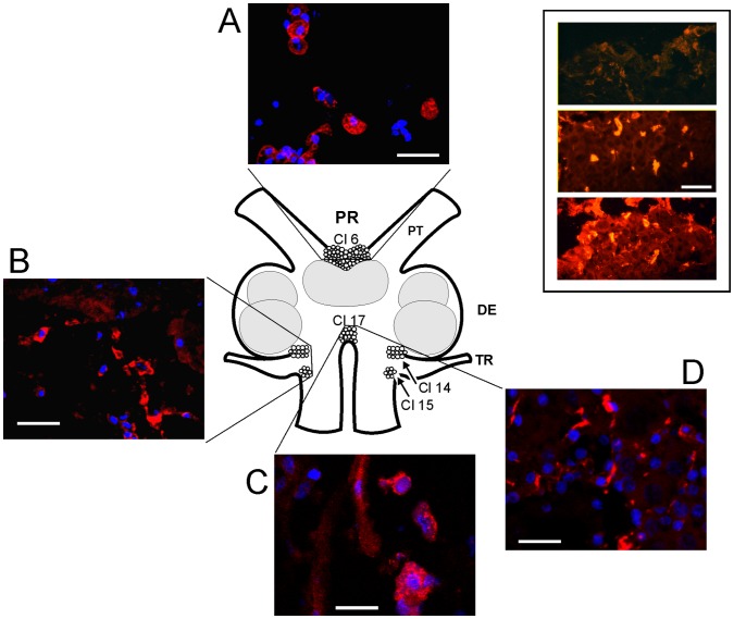
Figure 3. Confocal images showing brain CHH-positive cells and fibers.
Immunohistochemical localization of CHH-IR cells in the crayfish brain. The drawing represents a dorsal view of the brain as they relate to the representative confocal 1.6 µm optical sections stacked in 10 µm images of CHH-IR in the neuronal clusters (Cl 6, Cl 14,15 and 17). CHH is marked in red, and the cell nucleus marked in blue (DAPI). PR, protocerebrum; DE, deuterocerebrum; TR, tritocerebrum; PT, protocerebral tract. The white calibration bars equal 40 µm. The inset shows three horizontal serial sections of PR from the dorsal (upper panel) to central (lower panel) brain (CHH is shown in red). Immunopositivity is visible in all of the sections and is brighter in the central section. White calibration bar = 100 µm.

