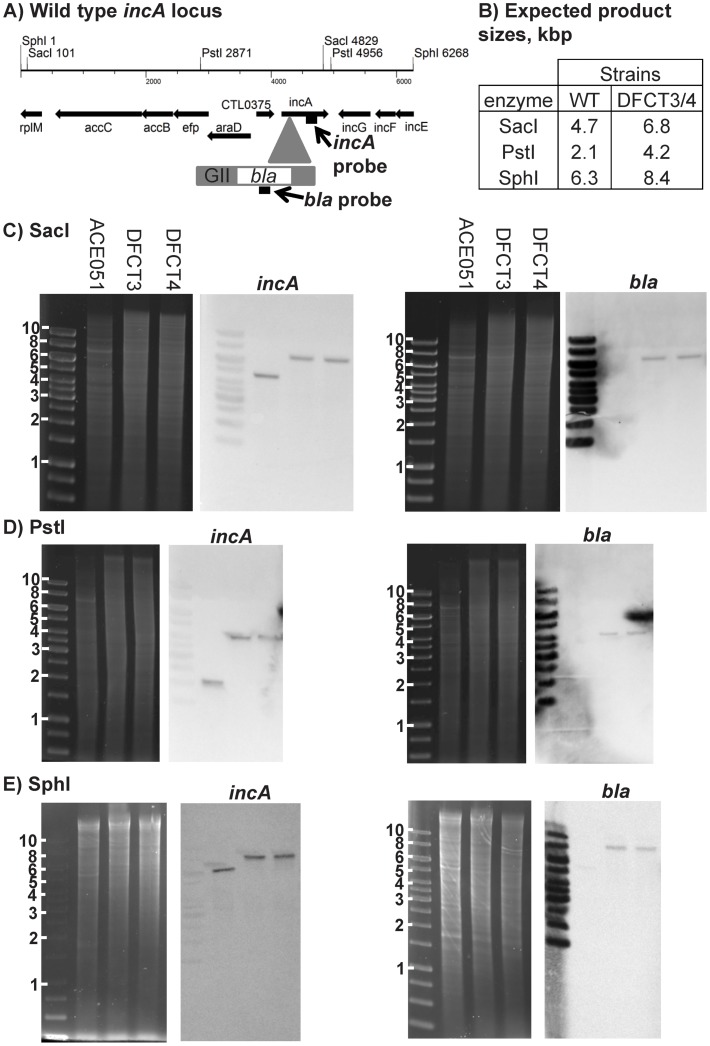
Figure 2. Southern blot characterization of intron insertion frequency in incA::GII(bla) clones.
Southern blotting was performed using DIG-labeled probes targeting the 3′ region of incA or the bla gene located within the intron. Probe locations, restriction site locations, and expected fragment sizes are shown in panels A and B, respectively. Genomic DNA was digested with either SacI, PstI, or SphI, separated on agarose gels, stained with ethidium bromide, and viewed under UV transillumination. DNA was then transferred to nylon membranes and probed with DIG-labeled incA or bla probes. Probes were detected using anti-DIG-antibodies conjugated to alkaline phosphatase and visualized with colorimetric substrate. DNA gels and their respective Southern blots are shown for each restriction enzyme used. Molecular weight markers and sizes (in kbp) are shown to the left of each DNA gel. DNA sources are shown at the top of each DNA gel and the probe used for the respective blot is shown above each blot.

