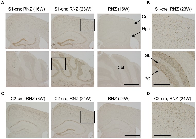
Figure 1. Detection of DNA recombination by synapsinI-cre or camk2a-cre transgene in mouse brain.
Transgenic mice for synapsinI-cre (S1-cre) or camk2a-cre (C2-cre) were crossed with RNZ mice. RNZ male mice harboring S1-cre or C2-cre at indicated weeks of age were subjected to anti-LacZ staining to detect cre-mediated DNA recombination. RNZ mice without a cre transgene were used as controls. (A) In S1-cre; RNZ mice at 16 weeks of age, LacZ-positive cells were strongly detected in dentate gyrus and CA3 in hippocampus and some cortical cells, but very few were seen in cerebellum. At 23 weeks, LacZ expression became wider in the cortex and hippocampus, and was clearly detected in cerebellum. No distinct LacZ expression was detected in the control RNZ mice. (B) High magnification of boxed region in (A). LacZ expression was broadly detected in multiple layers of cortex and Purkinje and granular cells of cerebellum in 23 week-old S1-cre; RNZ mice. (C) LacZ-positive cells were broadly detected in brains of 8-week-old C2-cre; RNZ mice, especially in layer II/III of cortex and CA1 of hippocampus. It became wider at 24 weeks of age. Again, no distinct LacZ expression was observed in the control RNZ mice. (D) High magnification of boxed region in (C), indicating detection of LacZ-positive cells in multiple layers of cortex in 24 week-old C2-cre; RNZ mice. Cor (cortex), Hpc (hippocampus), Cbl (cerebellum), PC (Purkinje cell) and GL (granular layer). Bars are 1 mm (A, C) and 0.4 mm (B, D).

