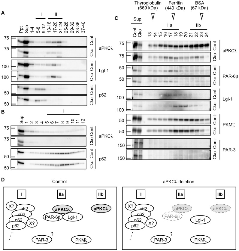
Figure 4. Gel filtration of cortical lysates of aPKCλ conditional deletion mice.
Cortex of 7-month-old female mice harboring aPKCλ flox/−; S1-cre (Cko) or flox/+ (Cont) was homogenized with lysis buffer. After centrifugation and removal of pellets (Ppt), supernatants (Sup) were subjected to gel filtration, and a total of 40 fractions were collected. Molecular weight markers were detected in Fr. 13–14 (669 kDa; thyroglobulin), Fr. 17–18 (440 kDa; ferritin), Fr. 23 (67 kDa; bovine serum albumin) and Fr. 29 (25 kDa; RNase). (A) Western blot analysis of Ppt, Sup, and mixture of four sequential fractions using antibodies for aPKCλ, Lgl-1 and p62. aPKCλ and Lgl-1 were detected mainly in fractions 13–24 (referred to as Fr. II) in the control cortex, whereas p62 was detected exclusively in fractions 5–12 (Fr. I). (B) Western blot analysis of Sup and fractions 1–12 using antibodies for aPKCλ and p62. p62 but not aPKCλ was highly detected in the Fr. I in control cortex. (C) Western blot analysis of Sup and fractions 13–24 using antibodies for aPKCλ, PAR-6β, Lgl-1, PKMζ (sc-216) and PAR-3. aPKCλ was broadly detected in fractions 15-24 in the control cortex, which could be separated into two fractions; Fr. IIa containing PAR-6β and Lgl-1, and Fr. IIb without containing aPKCλ-interacting proteins examined here. (D) Schematic model of potential protein compositions in the cortical lysates. In the control mouse, aPKCλ was incorporated into two major fractions: the Fr. IIa containing aPKCλ in a protein complex with PAR-6β and Lgl-1, and the Fr. IIb containing complex-free aPKCλ monomer. In contrast, aPKCλ was not clearly detected in the Fr. I containing large protein complex composed of p62 oligomer and some of its interacting proteins (indicated by an X). aPKCλ deletion induces reductions of aPKCλ in complex (IIa) as well as free aPKCλ (IIb), resulting in PAR-6β reduction and Lgl-1 dissociation from the complex.

