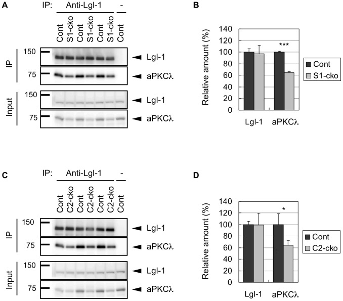
Figure 5. Immunoprecipitation assay using aPKCλ conditional deletion mouse brains.
(A) Cerebra of 11-month-old male mice harboring aPKCλ flox/flox (Cont; n = 3) or aPKCλ flox/flox; S1-cre (aPKCλ S1-cko; n = 3) were lysed (Input) and subjected to immunoprecipitation (IP) with anti-Lgl-1 antisera. IP without antisera (-) was used as a negative control. The input and IP samples were analyzed by Western blotting using antibodies for Lgl-1 and aPKCλ. (B) Bands of IP samples in (A) were quantified and plotted. (C) Cerebra of 20-month-old male mice harboring aPKCλ flox/flox (Cont; n = 3) or aPKCλ flox/flox; C2-cre (aPKCλ C2-cko; n = 3) were subjected to IP and analyzed as in (A). (D) Bands of IP samples in (C) were quantified and plotted. Note the significant reduction of aPKCλ co-immunoprecipitated with Lgl-1 in these aPKCλ deletion mouse cerebra. Values are means ±SD (*P<0.05, ***P<0.001).

