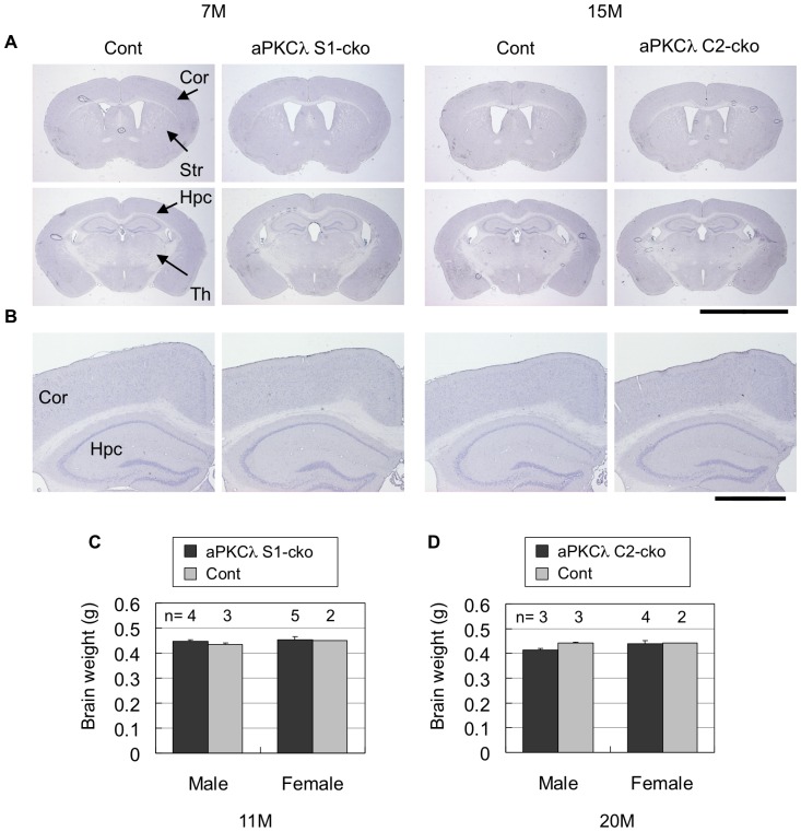
Figure 6. Hematoxylin staining and brain weights of aPKCλ conditional deletion mice.
(A) Hematoxylin staining of coronal sections of 7-month-old aPKCλ flox/−; S1-cre (S1-cko) or flox/+ (Cont) female mice (left two panels), or 15-month-old aPKCλ flox/flox; C2-cre (C2-cko) or flox/+; C2-cre (Cont) male mice (right two panels). (B) Magnified images shown in (A). (C, D) Brain weight of 11-month-old aPKCλ flox/flox (Cont) or flox/flox; S1-cre (S1-cko) male mice (C), or 20-month-old aPKCλ flox/flox (Cont) or flox/flox; C2-cre (C2-cko) male mice (D). Numbers of mice (n) used for analysis are indicated. Values are means ± SD. Cor (cortex), Str (striatum), Hpc (hippocampus) and Th (thalamus). Bars are 5 mm (A) and 1 mm (B).

