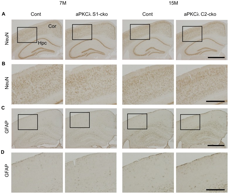
Figure 7. NeuN and GFAP staining of aPKCλ deletion mouse cerebrum.
Immunohistochemical analysis of 7-month-old aPKCλ flox/−; S1-cre (S1-cko) or flox/+ (Cont) female mice (left two panels), or 15-month-old aPKCλ flox/flox; C2-cre (C2-cko) or flox/+; C2-cre (Cont) male mice (right two panels). (A) Staining of coronal sections with anti-NeuN, a neuronal marker. (B) Magnified images of boxed regions shown in (A). No distinct reduction of NeuN-positive cells in these aPKCλ deletion mice was found. (C) Staining of coronal sections with anti-GFAP, an astrocyte marker. (D) Magnified images of boxed regions shown in (C). No distinct induction of astrogliosis in these aPKCλ deletion mice. Cor (cortex) and Hpc (hippocampus). Bars are 1 mm (A, C) and 0.4 mm (B, D).

