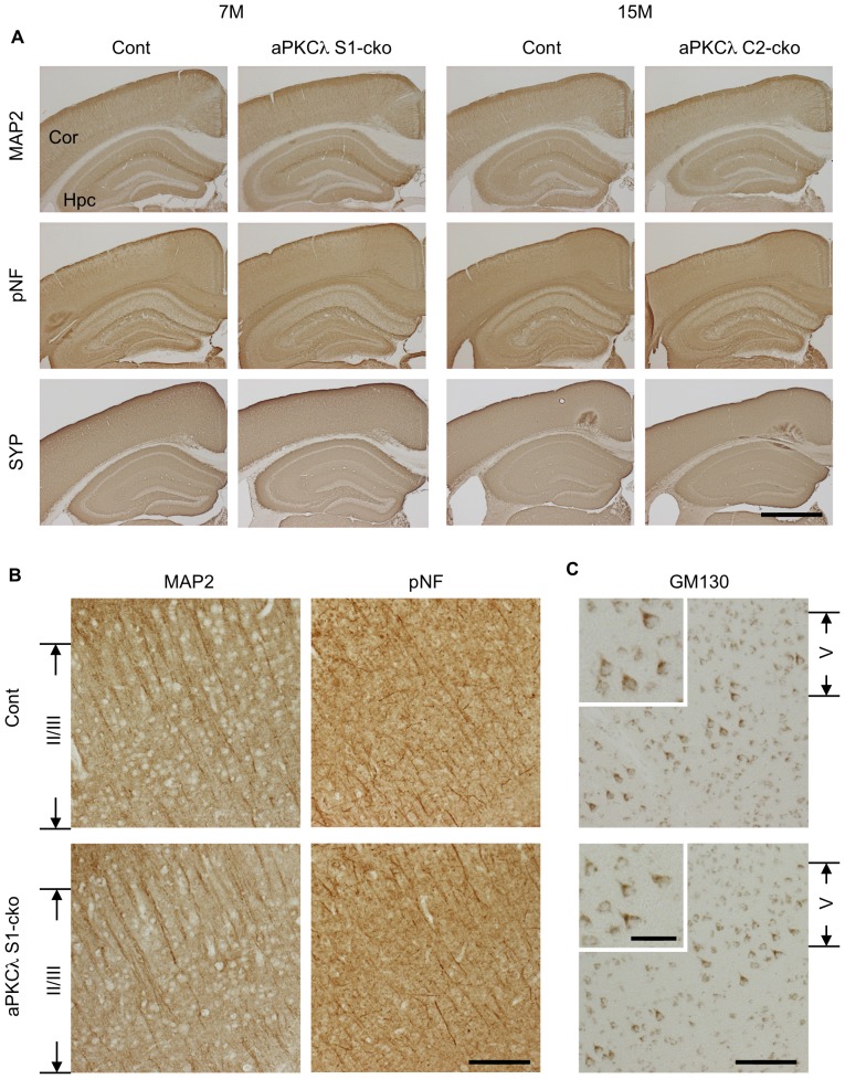
Figure 8. Neural marker staining of aPKCλ deletion mouse cerebrum.
Immunohistochemical analysis of 7-month-old aPKCλ flox/−; S1-cre (S1-cko) or flox/+ (Cont) female mice (left two panels), or 15-month-old aPKCλ flox/flox; C2-cre (C2-cko) or flox/+; C2-cre (Cont) male mice (right two panels). (A) Staining of coronal sections with antibodies for microtubule-associated protein-2 (MAP2), phospho-neurofilament (pNF) and synaptophysin (SYP), markers for dendrites, axons and synapses (pre-synapses), respectively. (B) Enlarged images for cortical layer II/III region of 7-month-old female mice stained with anti-MAP2 or anti-pNF antibody shown in (A). (C) Staining of coronal sections of 7-month-old female mice with antibody for GM130, a Golgi marker. Images for cortical layer V region are shown, and insets are enlarged images for layer V neurons. Note no distinct alteration in neuronal marker staining and Golgi location in aPKCλ deletion mice. Cor (cortex) and Hpc (hippocampus). Bars are 1 mm (A), 100 µm (B, C) and 40 µm (insets in C).

